4D Micro Computer Tomography
Methods FMF, Fiederle
| 4D Micro-CT | Model: | FMF model |
| Unit and Room: | Uniklinik, Nuklearmedizin, laboratory 5 | |
| Responsible: | Dr. M. Fiederle |
|
| Small Animal Computed Tomograph |
Further information: | www.fmf.uni-freiburg.de/service/ servicegruppen/sg_matchar/chat/ |
| Short Description: Investigations on pixelated X-Ray semiconductor detectors and on spectroscopic/energy selective X-Ray CT of small objects/animals with high resolution. |
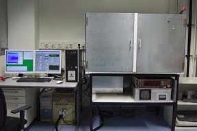
Picture of the Equipment
|
|
| Available Experiments/Techniques: High resolution X-Ray imaging of small objects: |
||
| Special Equipment: Microfocus X-Ray system Philips HOMX-161, energy 5 - 120 keV |
||
| Measurements on the equipment are currently done by: | Students after extensive training Trained scientific service personal |
|
| Recent Publications, where this instrument was important (citation): |
IEEE Transactions on Nuclear Science, Vol. 56, issue 4, pages 1795 - 1799. DOI 10.1109/TNS.2009.2025175 |
|
| Typical problems that may be solved with this instrument: | - Investigations on semiconductor X-Ray detectors - Investigations on 4D X-Ray imaging and tomography |
|
![]() 4D Micro Computer Tomography (this page as a pdf file)
4D Micro Computer Tomography (this page as a pdf file)


